Artificial intelligence (AI) is the most discussed topic today in medical imaging research, both diagnostic and therapeutic. For diagnostic imaging alone, the number of publications on AI has increased from about 100-150 per year in 2007–2008 to 1000–1100 per year in 2017–2018 and 1400-1500 in 2022-2023.
Apart from the popularity of Artificial Intelligence in medical imaging, It is a great technique to continue with because there are several tools and easy processes available.
Before AI, it was way too time-consuming for researchers to process medical images and find accurate results. But with Artificial Intelligence medical imaging, it gets better and faster. The predictions are more accurate and they help in better analysis of a patient’s condition.
Today, we will discuss the process and some tools that are used during Artificial Intelligence in medical imaging. So, let us get started with it.
Artificial Intelligence Medical Imaging Process
Object Detection
The first step of AI medical diagnosis online is acquiring the image from the available data from storage, database, or center, and an image is created in digital format. This image is generally captured through a general-purpose camera or real-time image-capturing tool such as MRI, CT-Scan, Radiology, Ultrasound, nuclear medicine imaging, etc.
Object Processing
The second step is to process the acquired image. The motive is to make it more clear and visible to identify shape, color, and texture by reducing noises and distortions.
You can understand the preprocessing stage in some steps, first to minimize the processing time acquired images are resized into a lower pixel resolution. Second, to retain and focus on the interested area, some image cropping is done so that unnecessary parts can be removed. Third, to eliminate the noise, filters are applied to the image. Last is to remove undesired color variation, the authentic RGB color is transformed into grayscale intensity.
Segmentation
The step of the process is the segmentation of the object image after resizing all pixels. The unnecessary part of the background is cropped to focus on the important area. All pixels with the same characteristics are stored in the same segment so that clarity of the image can be achieved.
Identification
The next step of the segmentation process is the feature identification of an object. The point is again the clarity of the image because different features of an image such as Asymmetry index, Entropy, Autocorrelation, Homogeneity, and Contrast are used for the classification stage. After extracting the feature of an object you can move on to the last step.
Classification
The last but not least step of Artificial Intelligence in Medical Imaging is classification. In this step, all extracted features are refined by selecting and classification methods. 70% of data is read and tested by several deep learning techniques such as K- Nearest Neighbor, Multi-Level perceptron, Decision Tree, Naïve Bayes, Logistic Regression, Support vector machine, Artificial Neural Networks (ANN), and Convolutional Neural Networks (CNN). This helps in clarifying the category of diseases at the supervisory stage.
The rest 30% of the data is used to predict the situation and whether the disease has been diagnosed or not.
Artificial Intelligence In Medical Imaging Application Tools
Scaida for Artificial Intelligence Medical Imaging
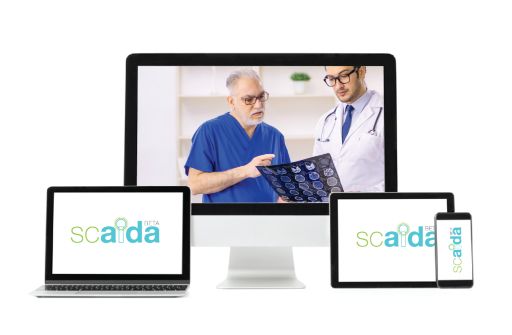
The very first tool on our list is Scaida, which is a medical imaging solution that allows healthcare organizations to store, view, and share medical images.
Key features of Scaida include a DICOM image viewer, cloud storage, and a patient image portal. The DICOM viewer allows staff to view X-rays, CT scans, MRIs, and other medical images. Images are stored securely in the cloud, with redundancy to prevent data loss. Patients can access medical images through the patient portal, providing ongoing access to images long after the visit.
Scaida Flow for Artificial Intelligence In Medical Imaging
Scaida Flow is a tool that integrates with any new legacy app, vendor, or platform. It supports multi-users from any location and device. It allows multi-users with zero footprints, image viewing, vendor document reading, scheduling, and patient portals. Overall, it is a great tool for smooth Artificial Intelligence In Medical Imaging.
Scaida Detect Artificial Intelligence Medical Imaging
It is a 100% cloud-based clinical trial imaging data detection tool. The workflow solution is designed to help cancer centers, trial sponsors, and CROs. It is helpful for those centers that have access to imaging RWD and better manage workflows, assessments, budgets, and audits.
Last Words
At the end of this blog, we can conclude that by using AI algorithms to analyze vast amounts of medical data and identify patterns and relationships, general AI medical diagnosis can transform the field of medicine. This can lead to improved patient outcomes and a more efficient and effective healthcare system in terms of AI in medical imaging.
ML Health is a great platform that can help you with better Artificial Intelligence In Medical Imaging through their impactful product and service. They have a clear vision to enhance the use of Artificial Intelligence Medical Imaging. For more information please check out the website today.


