-
AI Solutions
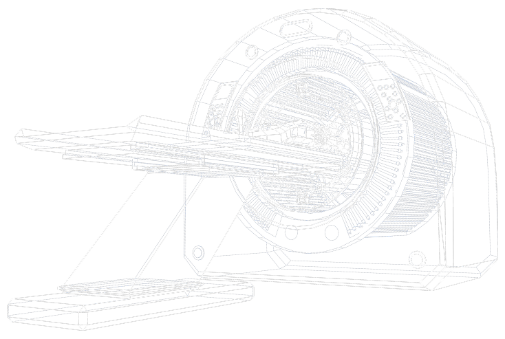
Imaging
WorkflowProvides a comprehensive, cloud-based
infrastructure for managing patient data,
imaging studies, and diagnostic
workflows—securely and efficientlyScaida
DetectCT
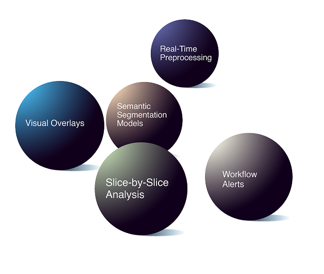
Accurately flags presence of anomalies in critical anatomical areas like the head, chest, abdomen, pelvis, and more.Scaida
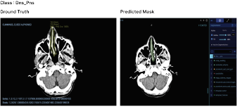
BrainCT
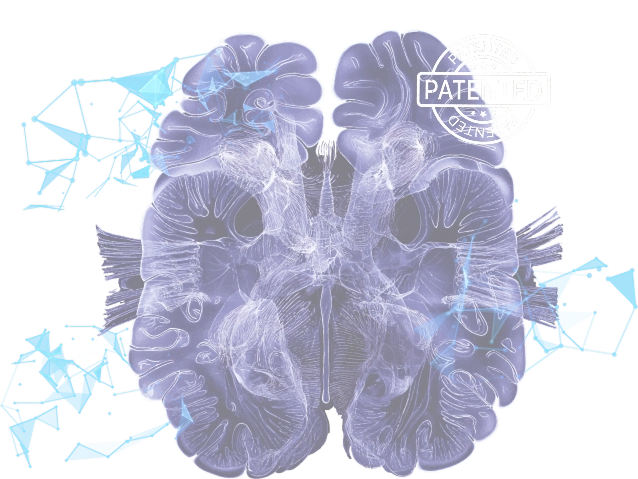
Provides precise brain segmentation and anomaly detection, assisting neuroradiologists in making life- saving decisions.Scaida
BrainCT-ICH
BrainCT-ICH identifies various types of hemorrhages using a slice-by-slice analysis model.NeuroCatch®
PlatformComplements your imaging data by quantifying cognitive function at the point of care. It provides a functional baseline alongside your structural diagnosis.❯ - Media
- Careers